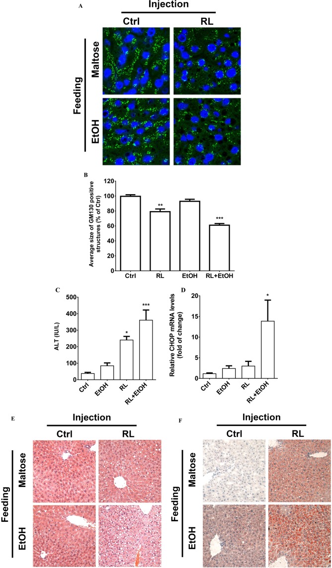
Figure 4.

Association of Golgi fragmentation with liver injuries in mice injected with anti‐HIV drugs and/or fed with alcohol. (A) Confocal images (×1000) showing Golgi fragmentation of hepatocytes of liver sections. The Golgi morphology was probed with immunofluorescence (green) using anti‐GM130 antibodies. The nuclei of hepatocytes were revealed with Hoechst blue staining. Ctrl, sample from mice injected with vehicle control and fed with isocaloric control diet; RL, mice injected with RTV+LPV and fed with control diet; EtOH, sample from mice injected with vehicle control and fed with alcohol diet. The image at the lower right corner represents samples from mice injected with RL and fed with an alcohol diet (i.e., RL+EtOH). (B) Quantitation of the Golgi fragmentation. (C) Levels of serum ALT in mice treated with the drugs and/or alcohol. (D) CHOP expression in liver samples from RL‐injected mice with or without alcohol feeding. (E) H&E staining of liver tissues showing fatty liver injury (×200). (F) Oil Red O staining of liver tissues showing fat accumulation in mice treated with the drugs and/or alcohol (×200). *P < 0.05; **P < 0.01; ***P < 0.005 compared to control; n = 4‐6. Error bars indicate standard error mean (SEM). Abbreviation: EtOH, ethanol.
