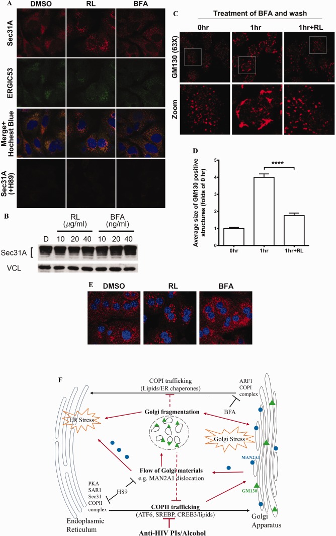
Figure 6.

Effects of anti‐HIV drugs on ER–Golgi trafficking. (A) Accumulation of COPII in the drug‐treated HepG2 cells and inhibition of COPII assembly by H89. The COPII complex was revealed by immunofluorescent staining with anti‐Sec31A antibody as shown in the confocal images (red). Anti‐ERGIC53 antibody was used to label the ERGIC structure by immunofluorescent staining (green, ×630). (B) Expression of Sec31A protein in RL‐ and BFA‐treated HepG2 cells. Cells were treated with RL or BFA at different concentrations for 8 hours. (C) Recovery of Golgi morphology after removal of BFA, which was inhibited by RL (×630). The cells were treated first with BFA (100 nM) for 1 hour and then treated with RL for 1 hour after BFA had been removed from the media. (D) Quantitation of Golgi fragmentation. (E) Accumulation of COPII complex in PMH treated with DMSO, RL, and BFA. COPII was probed with immunofluorescence (red) using anti‐Sec31 antibodies (×630). (F) Diagram depicting sites of the ER‐to‐Golgi trafficking targeted by the drugs. The red lines depict that the drugs disrupt COPII trafficking, causing Golgi stress, Golgi fragmentation, enzyme dislocation/redistribution, and ER stress. Error bars indicate standard error mean (SEM). Abbreviations: ARF1, ADP‐ribosylation factor 1; DMSO, dimethyl sulfoxide; ERGIC, ER–Golgi intermediate compartment.
