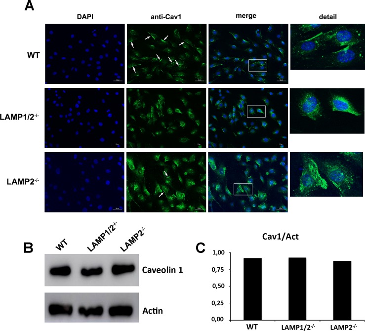
Fig 6. Absence of LAMP leads to decrease in caveolin associated with cell plasma membrane.
(A) WT, LAMP1/2-/-, or LAMP2-/- cells were fixed, submitted to labeling with anti-caveolin 1 and analyzed in a fluorescence microscope. Arrows indicate the presence of caveolin-1 at the cell surface. Details of each image are shown on the side. (B) WT, LAMP1/2-/- or LAMP2-/- cells were submitted to total protein extraction. Extracts were run on a gel, blotted onto nitrocellulose membranes and revealed using an anti-caveolin 1 antibody. The panel shows the presence of caveolin in the protein extracts from all three cells. (C) Graph shows the quantification of the amount of total caveolin in the different cell types.

