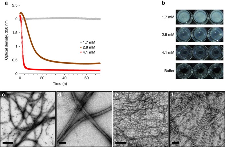
Figure 1. Self-assembly of the 20-mer peptide into ordered nanofibres.
The nanofibres formed in phosphate buffer. (a) Kinetics of absorbance at 350 nm at three concentrations over a period of 72 h and (b) macroscopic visualization of the preparations at the end of the experiment. (c,d) TEM micrographs of the 20-mer nanofibres at 1.7 mM or (e,f) 4.1 mM. Scale bars in c and e are 500 nm; scale bars in d and f are 100 nm.

