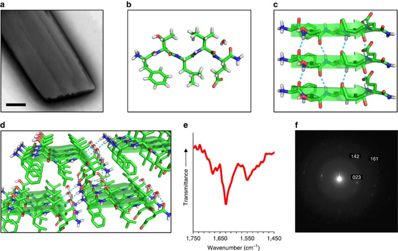
Figure 4. Structure of the 5-mer (FTLIE) microcrystals.
(a) TEM micrograph of an individual microcrystal grown in phosphate buffer at a concentration of 2.9 mM. (b) View of the asymmetric unit as determined for 5-mer single-crystals by XRD, showing a single peptide molecule and a water molecule. (c) Crystal packing of three peptide molecules along the crystallographic a axis. The peptide molecules are organized as a supramolecular parallel β-sheet. (d) Extended crystal packing, showing a network of hydrogen bonds and electrostatic interactions between β-sheets along the crystallographic b and c axes. For panels c,d, hydrogen bonds and electrostatic interactions are shown as dashed cyan and black lines, respectively. (e) FTIR spectrum of microcrystals grown in phosphate buffer at a concentration of 11.6 mM. (f) SAED pattern of an individual microcrystal grown in buffer at a concentration of 16 mM. Reflections are indexed in relation to the single crystal structure as obtained by XRD. Scale bar in a is 500 nm.

