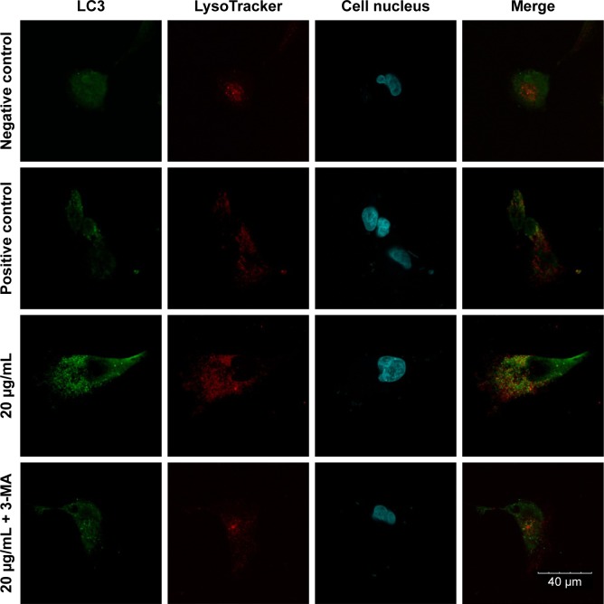
Figure 4.
Confocal immunofluorescence images.
Notes: The MC3T3-E1 cells were cultured with 20 μg/mL Ta-NPs for 24 h, with or without pretreatment with 3-MA (10 mM) for 1 h; cells treated with α-MEM alone served as a negative control group, and cells treated with 200 nM rapamycin served as a positive control group. The LC3-labeled green puncta (a characteristic of autophagosomes) could be clearly observed in the positive control group and in the 20 μg/mL Ta-NP-treated group but were nearly indistinguishable in the negative control group. When cells were pretreated with 3-MA, the amount of green puncta decreased sharply. Experiments were repeated three times. Scale bar: 40 μm. Magnification ×1,200.
Abbreviations: 3-MA, 3-methyladenine; α-MEM, alpha-modified Eagle’s medium; Ta-NPs, Ta nanoparticles.

