
Fig. 1.
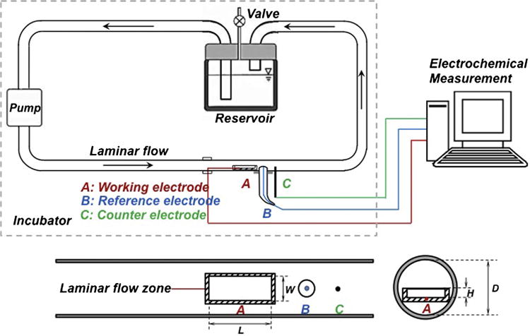
Schematic of the vascular bioreactor with an electrochemical monitoring system and close-up upside view of the test channel with the cross section view of specimen in place. Note: L = length of the specimen; W = width of the specimen; H = height of the specimen; D = inner diameter of test channel.