
Fig. 5.
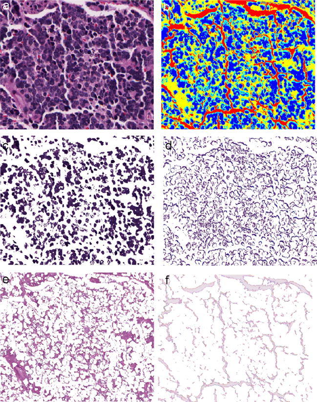
The segmented components in a typical image from undifferentiated subtype are shown. (a) Original image. (b) Partitioned image shown in colors with nuclei in blue, cytoplasm in cyan, neuropil in yellow, red blood cells in gray, and background in red. (c) Nuclei component. (d) Cytoplasm component. (e) Neuropil component. (f) Background component.