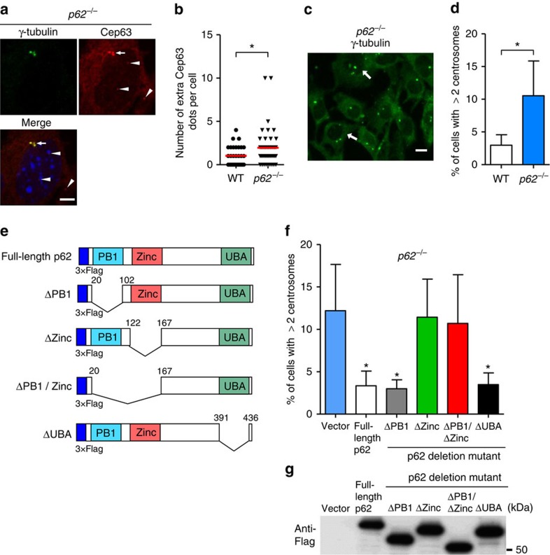
Figure 5. Involvement of p62 in centrosome number regulation.
(a,b) Increase in extra Cep63 dots in p62−/− MEFs. p62−/− MEFs were immunostained with anti-γ-tubulin and anti-Cep63 antibodies and were examined by fluorescence microscopy. Representative images of γ-tubulin (green), Cep63 (red) and the merged image are shown in a. The nucleus was stained with 4,6-diamidino-2-phenylindole (DAPI) (blue). Arrow and arrowheads indicate mature centrosome and extra Cep63 dots, respectively. Scale bar, 5 μm. (b) The number of extra Cep63 dots per cell was calculated (n>30 cells). The red lines indicate mean values. The asterisk indicates a significant difference (P<0.05, Student's t-test). (c,d) Increase in mature centrosomes in the p62−/− MEFs. The p62−/− MEFs were immunostained with anti-γ-tubulin antibody and were examined by fluorescence microscopy. A representative image is shown in c. Arrows indicate cells with multiple centrosomes. Scale bar, 10 μm. (d) The percentage of cells with three or more centrosomes is shown as the mean+s.d. (n=3). The asterisk indicates a significant difference (P<0.05, Student's t-test). (e) The design of p62 mutant plasmids. The p62 protein contains PB1, zinc-binding and UBA domains. Each deletion plasmid was generated. (f) The p62−/− MEFs were transfected with each Flag(3 × )-tagged plasmid and were immunostained with anti-γ-tubulin antibody. The percentage of cells with three or more centrosomes was calculated. Data are mean+s.d. (n=3). *P<0.05 versus the value of vector (analysis of variance (ANOVA) Tukey's post hoc test). (g) The equal expression of each p62 mutant was confirmed by western blotting using anti-Flag antibody. Uncropped images are shown in Supplementary Fig. 14.

