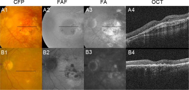
Figure 3.

Cases that mimic geographic atrophy (GA). (A1–A4) The 4-year follow-up images of a 51 year-old male participant with active choroidal neovascularization are depicted. Findings of GA include: color fundus photography (CFP; A1) showing a central patch of what appears to be depigmentation with sharply demarcated borders and visibility of underlying choroidal vessels; fundus autofluorescence (FAF; A2) showing hypo-autofluorescence in an area of apparent depigmentation; and late fluorescein angiography (FA; A3) showing hyper-fluorescence bounded by sharp borders. However, optical coherence tomography (OCT; A4) shows fibrous proliferation at the level of the retinal pigment epithelium (RPE) with associated shadowing, making classifying the depigmented lesion as GA problematic. (B1–B4) The 3-year follow-up images of a 69 year-old male participant with active choroidal neovascularization are depicted. Findings suggestive of GA include: FA (B2) showing hypo-autofluorescence with sharp borders in areas corresponding to patchy hypopigmentation on CFP (B1); and late FA (B3) showing corresponding hyper-fluorescence bounded by sharp borders. However, OCT (B4) shows irregular thickening at the level of the RPE with associated shadowing, also making classifying the depigmented lesion as GA problematic.
