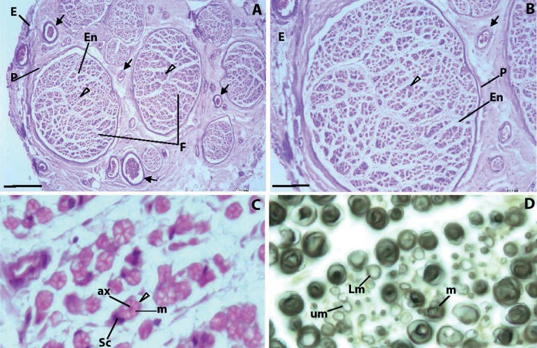
Figure 1.
Typical appearance of peripheral nerves. A, B. Each nerve is surrounded by a dense connective tissue, the epineurium (E). Nerves are a group of nerve fibers (arrowheads) disposed into different size aggregates - the nerve fascicles (F). Nerve bundles are separated by loose connective tissue, the perineurium (P). Numerous blood vessels (arrows) can be seen in the perineurium. Each bundle presents numerous axons surrounded by loose connective tissue, the endoneurium (En). In myelinated axons, Schwann cells develop thin cytoplasmic extensions that wrap over the axon surface, creating the myelin sheath. The cytoplasmic membrane of the Schwann cell is tightly connected to the axon cytoplasmic membrane, controlling axon metabolism through transmembrane channels and pH, providing the ATP necessary for kinesin-continued transport of neurotransmitter vesicles along microtubules, and scavenging free toxic oxygen radicals produced during electric impulses. The myelin layer is a barrier to most molecules, and only very small liposoluble molecules can traverse the myelin sheath. C. In myelinated axons, each nerve fibber consists of an axon (ax) surrounded by the myelin sheath (m) of Shawn cells (Sc). Unmyelinated axons are nevertheless surrounded by Schwann cells. D. Myelinated fibers (m), light myelinated fibers (Lm) and unmyelinated fibers (um). A-C. Sural nerve, Hemalumen-eosin. D. Sciatic nerve, Osmic acid. Bars: A: 200 µm; B: 100 µm; C: 9 µm; D: 9 µm.

