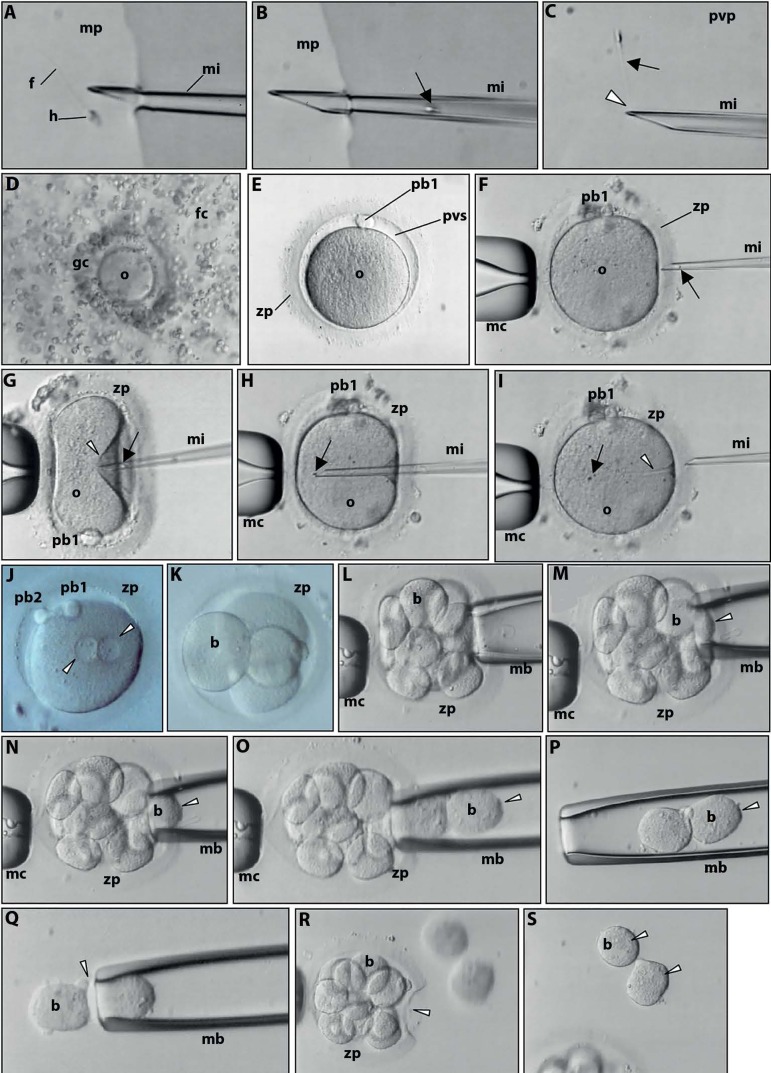
Figure 3.
Live images observed at the inverted microscope. Preimplantation genetic diagnosis. A. Selection of a single, morphologically normal spermatozoon (head-h; tail-f) with a 3D forward progressive motility in culture medium (mp) supplemented with PVP to decrease spermatozoon velocity. Microinjection pipette (mi). B. Aspiration of the selected spermatozoon (arrow) into the microinjection pipette (mi). C. Immobilization of the selected spermatozoon (arrow) in PVP (pvp) by crashing (arrowhead) the distal microtubules of the flagellum. D. Cumulus-oocyte complex of the aspirated ovarian follicle. The oocyte (o) is coated by granulosa cells (gc) and encircled by follicular cells (fc) of the cumulus oophorus. E. Mature metaphase II oocyte after denudation of the cumulus and granulosa cells. Note the perivitelline space (pvs), the first polar body (pb1) and the glycoprotein oocyte coat, the zona pellucida (zp) F. For microinjection, the oocyte is held by a contention micropipette (mc) with the first polar body at 3 (F) or 6 (G) o’clock. The microinjection pipette is penetrating the zona pellucida. The spermatozoon is at the distal border of the microinjection pipette, head first (arrow). G. Penetration of the oocyte membrane (arrowhead) by the microinjection pipette. H. The spermatozoon (arrow) is released in the ooplasm. I. Extrusion of the microinjection pipette, leaving the spermatozoon in the ooplasm (arrow). The furrow of the oocyte is still visible (arrowhead). J. The day after microinjection the oocyte shows signs of normal fecundation as revealed by the presence of the second polar body (pb2) and of both pronuclei (arrowheads). K. At the second day after microinjection the zygote cleaved into an embryo with 4 blastomeres (b). L-P. Successive images of the embryo biopsy. Three days after microinjection the embryo has 8 blastomeres. After opening a small hole in the zona pellucida, the embryo biopsy pipette (mb) enters the perivitelline space and gently aspirates two blastomeres (arrowhead). Q. Extrusion (arrowhead) of the two blastomeres to the surrounding medium. R. The embryo is left in culture with the evident zona pellucida hole (arrowhead). S. Note the nuclei of the isolated blastomeres (arrowheads). The diameter of the oocyte is about 110 µm.

