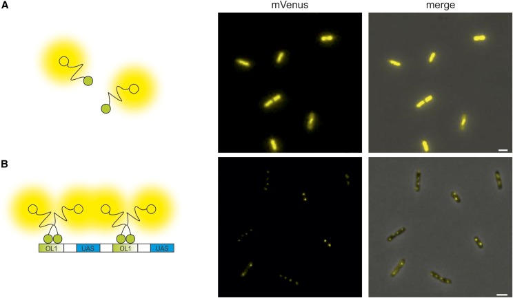
Figure 2.
Specific binding to a hybrid FROS array. Fluorescence microscopy images of cells without (A, strain SM69) and with (B, strain SM77) OL1/UAS hybrid FROS array expressing a fusion of the λcI repressor to full-length mVenus. Left panel presents cartoons illustrating fluorescent fusion proteins in respective E. coli strains. Fluorescence microscopy images show the mVenus channel (middle panel) or merges of mVenus and phase-contrast channel (right panel). Exposure time was set to 100 msec. Bar, 2 µm.

