Figure 1.

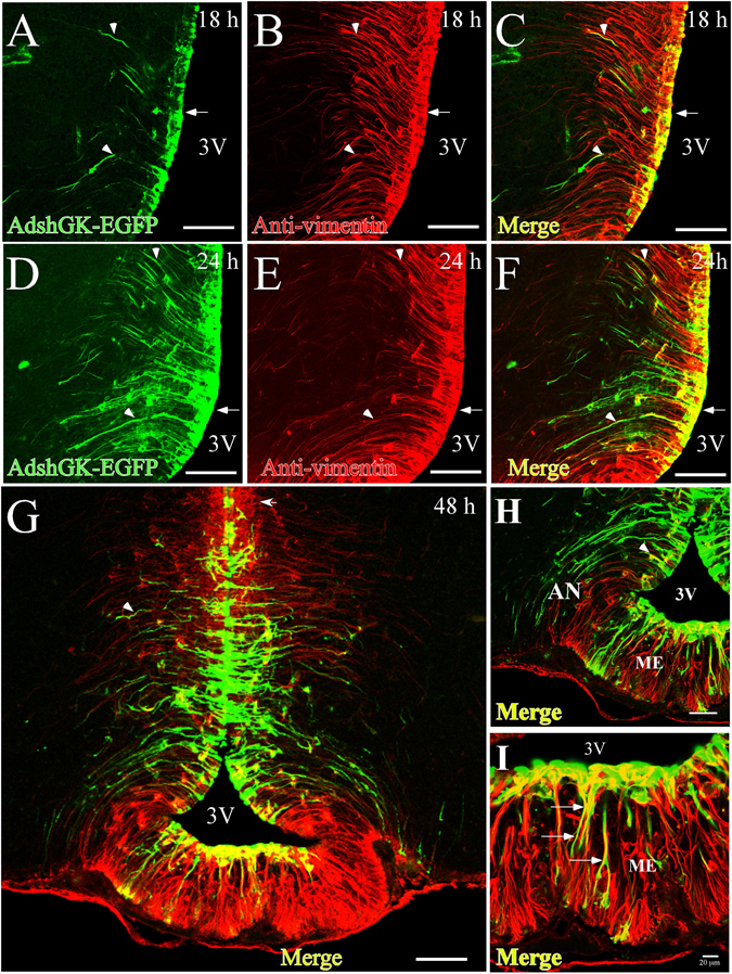
Time-course of adenoviral transduction in the basal hypothalamus. (A–L) Frontal sections (40 µm) from the hypothalamus at 18 h (A–C), 24 h (D–F) and 48 h (G–I) following injection of adenoviral particles (Ad-shGK-EGFP) into the 3V. EGFP fluorescence (green) and immunoreactivity for vimentin (red) and colocalization of EGFP and vimentin (yellow) are shown. Arrows show the processes of transduced tanycytes. (G) Panoramic view of the basal hypothalamus, showing tanycyte transduction. (I) Higher magnification image showing that Ad-EGFP transduction occurs in β2-tanycytes. AN, arcuate nucleus; 3V, third ventricle; ME, median eminence. Scale bars (A–F and H) 50 µm, (G) 100 µm, (I) 20 µm.
