
Figure 4.
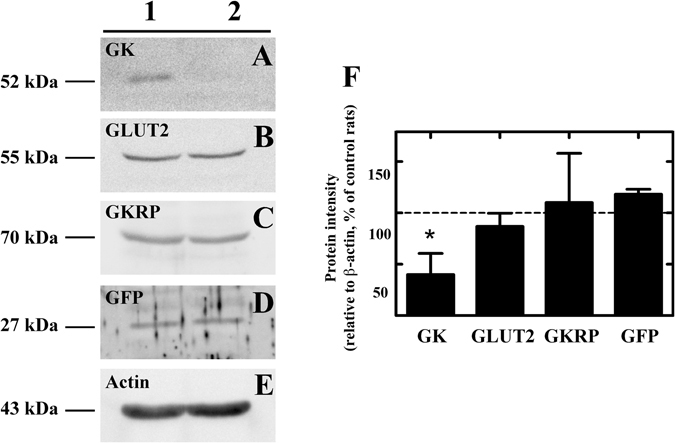
GK inhibition 48 h after AdshGK injection into the 3V. (A) Western blot assays of hypothalamic protein extracts in control (Ad-shβGal-injected rats) and Ad-shGK-injected rats using anti-GK, anti-GLUT2, anti-GKRP, and anti-GFP (transduction control) antibodies. An anti-β-actin antibody was used as loading control. Images are representative of three different experiments. (B) Densitometric analysis of each protein relative to β-actin evaluated in Ad-shGK-injected rats, as a percentage of this protein in Ad-shβgal-EGFP-injected rats. Statistics: t-test with Welch correction *p < 0.05.