
Figure 2.
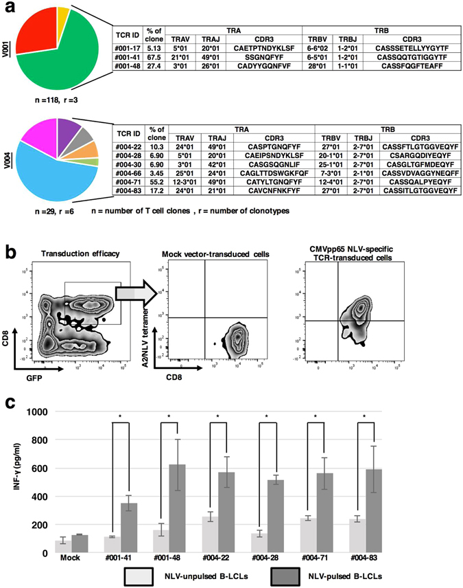
Characterization of CMV NLV-specific TCRα and TCRβ repertoires of single cells. (a) CMV NLV-specific TCRα and TCRβ repertoires identified using single-cell multiplex clonotypic analysis of two HLA-A2-positive and CMV-seropositive healthy donors (V001 and V004). Single CMV NLV-specific T cells were sorted into 96-well PCR plates and then cDNAs were amplified using multiplex RT-PCR. The PCR products were sequenced and conceptually translated to identify CDR3α and CDR3β. We analysed 118 and 29T cells from V001 and V004, respectively, and identified 3 (TCR ID; 001-17, 48 and 41) and 6 (TCR ID; 004-66, 22, 63, 30, 28 and 71) CDR3α and CDR3β pairs from each respective donor. (b) Transduction of sequences encoding CMV NLV-specific TCRs. TCRα and TCRβ pairs were cloned into expression vectors encoding GFP that were used to transfect PHA blasts derived from a CMV-seronegative healthy donor. Representative flow cytometric profiles illustrate transduction efficiencies. Cells with double-positive staining for the NLV tetramer and GFP were considered successful transductants. (c) IFN-γ production by TCR-transduced cells. TCR-transduced cells were co-cultured for 16 h with NLV-pulsed and NLV-unpulsed B-LCLs derived from the cognate donors. The IFN-γ concentrations in culture supernatants were measured using an IFN-γ ELISA. The results represent the mean ± standard deviation (S.D.) of triplicate experiments. The error bars represent standard deviation. The asterisks indicate significant difference (p < 0.05) in the IFN-γ concentrations between samples co-cultured with NLV-pulsed and NLV-unpulsed B-LCLs (paired t-test). Although mock-transduced PHA blasts did not recognize NLV peptide-pulsed or control B-LCLs, all TCR-transduced PHA blasts recognized only NLV peptide-pulsed B-LCLs, verifying the antigenic specificity of the TCR-transduced PHA blasts.