
Figure 1.
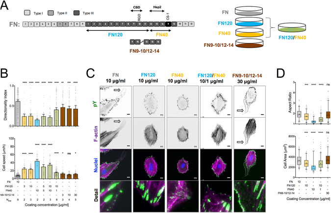
Signals from both the cell binding and C-terminal heparin-binding domains of FN are required for high directional persistence in fibroblast migration. (A) Schematic representation of plasma FN, showing the location of the different domains, the proteolytic fragments and recombinant fragments relevant for this study. Substrates were coated with FN, FN fragments or their combinations (color coded). (B) Cell speed and directionality index (distance from the origin divided by the trajectory length) were calculated for single REF migrating on indicated substrates for 16 hours. Cell speed was the lowest and directionality index was the highest on FN-coated substrates. Nexp: number of independent experiments. Mean ± s.e.m. are presented. (C) Epifluorescence microscopy images of REF seeded for 6 hours on indicated substrates revealed important differences in adhesion cluster formation and F-actin cytoskeletal organization (see main text for details). Polarized protrusions are indicated by block arrows on FN and FN9–10/12–14. (D) REF projected cell area and aspect ratio, 6 hours post-seeding on substrates coated with FN or FN fragments. Results from >150 cells and 2 independent experiments are presented. The middle line in box plots indicates the median, the box indicates the interquartile range, the whiskers the 5th and 95th percentiles and the cross the mean. Scale bars: 10 μm. Data in (B,D) were compared to control FN-coated substrates using one-way ANOVA analysis. ns: not significant; *P < 0.05, **P < 0.01, ***P < 0.001, ****P < 0.0001.