Figure 7.

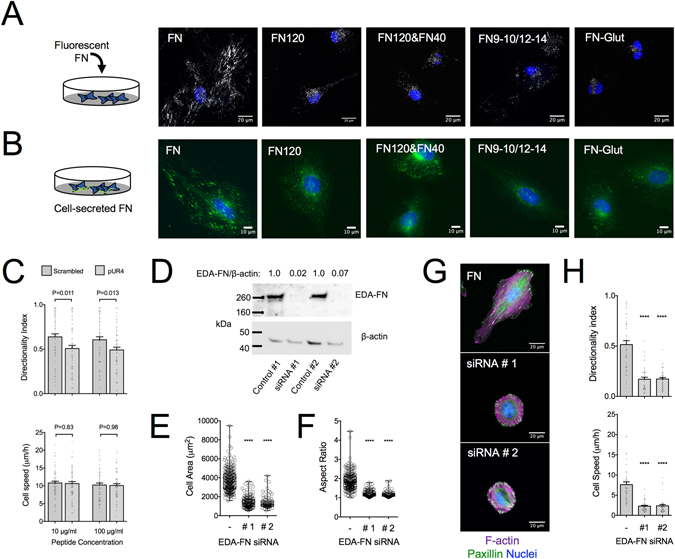
FN fibrillogenesis inhibition impairs directional persistence in fibroblast migration and cellular FN knockdown abolishes migration altogether. (A) Confocal microscopy images of fluorescently-labeled FN (gray) bound and/or assembled by REF on substrates with indicated coatings. Culture medium was exchanged 4 hours after seeding with medium containing 10 μg/ml FFN and incubated for another 20 hours, prior to washing and cell fixation. Fibrillogenesis of soluble FFN was observed only on FN-coated substrates. Nuclei are stained with DAPI (blue). (B) Epifluorescence microscopy images of REF immunostained against cellular FN (green), fixed 24 hours after seeding on substrates with indicated coatings. FN fibrillogenesis was evident only on FN-coated substrates. Nuclei are stained with DAPI (blue). Coating concentrations of FN: 10 μg/ml, FN120: 10 μg/ml, FN120/FN40: 10/1 μg/ml and FN9–10/12–14: 10 μg/ml. (C) REF directionality index on FN-coated substrates was reduced in presence of pUR4 peptide compared to a scrambled control peptide, whereas cell speed was unaffected. Values were compared using a Mann-Whitney test (Nexp = 3; n = 60). Mean ± s.e.m. are presented. (D) Western blot analysis of lysates from REF treated 48 hours with siRNA against EDA-FN. The EDA-FN/β-actin ratio was obtained by densitometric analysis. (E) REF projected cell area and (F) aspect ratio 6 hours post-seeding on substrates coated with FN. EDA-FN knockdown resulted in a large reduction of cell area and cell rounding (Nexp = 2). Values of siRNA-treated REF were compared to the control condition using one-way ANOVA analysis (****P < 0.0001). (G) Epifluorescence microscopy images of control or siRNA-treated REF seeded for 6 hours on FN-coated substrates, fixed and stained against paxillin (green), F-actin (magenta) and DNA (blue). EDA-FN knockdown changed dramatically cell morphology but did not impair focal adhesion or stress fiber formation. (H) REF speed and directionality index calculated for control and siRNA-treated REF on FN. EDA-FN knockdown abolished migration, with residual cell speed reflecting nuclei movements within the cell body. Data were compared using one way ANOVA analysis (****P < 0.0001). Mean ± s.e.m. are presented (Nexp = 2; n = 40).
