
Figure 1.
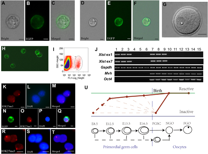
The X chromosome undergoes a process of reactivation in female germ cells from FGSCs to oocytes. (A–C) Representative morphology of Mvh positive cells from XX genital ridges after E12.5 under light microscopy and fluorescence microscopy. (D–F) Representative images of Mvh positive cells from ovaries of 3–5 days old Mvh-GFP transgenic mice (Mvh-cre; ROSAmT/mG mice) under light microscopy and fluorescence microscopy. (G) Representative morphology of oocytes. (H) Representative view of merger for Mvh positive cells under light microscopy and fluorescence microscopy after purification with FACS.(I) An example for FGSC purification with FACS. (J) Single cell RT-PCR analysis of Xist, Mvh and Oct4. Lane 1–3, female Tail fibroblasts (positive control); lane 4–6, male tail fibroblasts (negative control); lane 7–9, FGSCs; lane 10–12, NGO; lane 13–15, FGO. Full-length gels are presented in Supplementary Fig. S15. (K–M) FGSC cell line showed the expected frequencies of the Xi-like enrichment for H3K27me3. Cells were counterstained with DAPI. (N–Q) Fresh isolated FGSCs showed the expected frequencies of the Xi-like enrichment for H3K27me3. Cells were counterstained with DAPI. (R–T) Female tail fibroblasts showed the expected frequencies of the Xi-like enrichment for H3K27me3 (positive control). Cells were counterstained with DAPI. (U)Schematic illustration of the process of reactivation in germ cell development: the FGSCs, PGCs and oocytes formed a process from X inactivation to X reactivation in germ cell development. The solid line shows the process we determined, while the dotted line shows the process we do not test. The slim dotted line indicated that early germ cells contain the cells entering meiosis and very small amount of the cells not entering meiosis (or precursors of FGSCs). For (A–F,H,K–T), Scale bar = 5 μm, for G, Scale bar = 20 μm. The experiments were conducted three times.