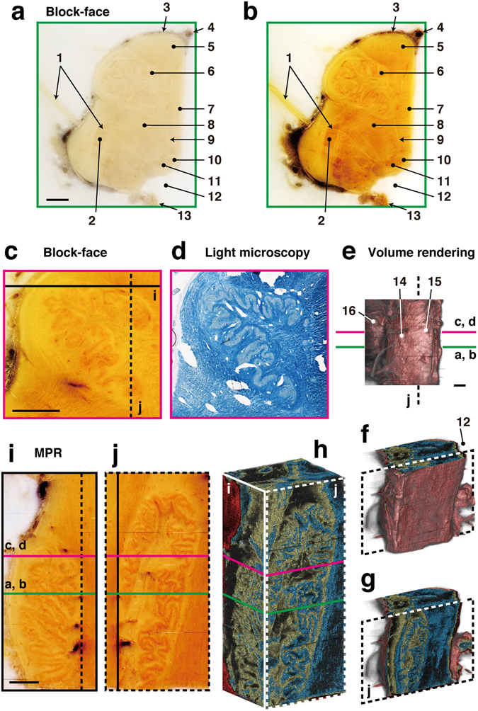
Figure 4.

Correlative 2D and 3D images of human brainstem. The right half of the brainstem was cut into sections at a thickness of 20 µm. Volume rendering and MPR images were reconstructed from 1275 block-face images. (a) A block-face image taken in the horizontal plane is shown as observed by the naked eye. (b) The image was processed to enhance the contrast between nerve nuclei and fibers. (c,d) A block-face image was correlated with a section stained with LFB to confirm the nerve nuclei and fibers. (e–h) Volume rendering shows the surface of the brainstem (e,f), and the folds in the olivary nucleus (g,h). (i,j) MPR images show real-color images of the olivary nucleus in the coronal plane (i), and the sagittal plane (j). 1: vagus nerve rootlets, 2: spinal nucleus of trigeminal nerve, 3: pia matter, 4: basilar artery, 5: pyramidal tract, 6: principal olivary nucleus, 7: medial lemniscus, 8: reticular formation, 9: hypoglossal nerve rootlets, 10: hypoglossal nucleus, 11: dorsal vagal nucleus, 12: fourth ventricle, 13: choroid plexus, 14: olive, 15: pyramid, 16: pons. Bars: 2 mm.
