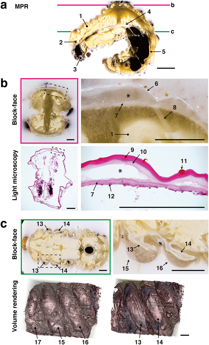
Figure 5.

Correlative 2D and 3D images of a stag beetle larva. A whole stag beetle larva was cut into sections at a thickness of 20 µm. MPR and volume rendering were reconstructed from 1064 block-face images. (a) The sagittal plane was reconstructed by MPR. (b) Sections were stained with H&E, and correlated with block-face images. The gap between the newly developing pupal cuticle and the old larval cuticle was maintained in both the block-face image and the section (asterisk). (c) Two pairs of wings are shown in the block-face image. Volume rendering shows the surface structures of the thorax, and a forewing and hindwing within the thorax. 1: fat body, 2: brain, 3: oral cavity, 4: midgut, 5: hindgut, 6: larval cuticle, 7: pupal cuticle, 8: muscle, 9: larval exocuticle, 10: larval endocuticle, 11: hair, 12: epidermis, 13: forewing, 14: hindwing, 15: mesothorax, 16: metathorax, 17: prothorax. Bars in a: 5 mm, b, c: 1 mm.
