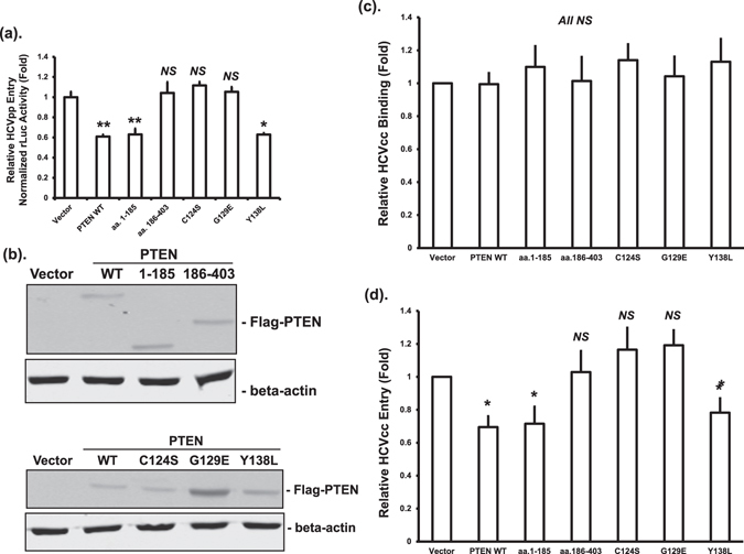
Figure 2.

PTEN inhibits HCV entry through its lipid phosphatase activity. (a) Huh-7.5 cells were transfected with plasmids expressing wild-type Flag-PTEN (PTEN WT), truncated mutants, phosphatase deficient mutants, or empty vector. Forty eight hours after transfection, cells were infected using HCV-2a J6 pseudoparticles with a firefly luciferase reporter (HCVpp-Luc). Luciferase activity was measured 24 hours after infection and normalized to total protein amount. Luciferase activities were expressed as fold changes relative to vector control. Statistical differences between samples were demonstrated as * if p ≤ 0.05, ** if p ≤ 0.01, or NS for not significant. This experiment was performed three times. (b) The protein levels of Flag-tagged PTEN after transfection were determined by Western blotting using an anti-Flag antibody. The β-actin levels were determined by blotting cell lysates with a β-actin antibody as loading controls. (c and d) Huh-7.5 cells were transfected with plasmids expressing wild-type PTEN (PTEN WT), truncated mutants, phosphatase deficient mutants, or empty vector, and infected with HCV-2a J6 core-Flag/JFH-1 (p7-rLuc-2A) virus 48 hours after transfection. After incubation at 4 °C for 1 hour (c, virus binding only) or after a further incubation at 37 °C for 3 hours (d, post-binding entry), cells were harvested and RNA extracted. HCV RNA levels were quantified by real-time PCR. Statistical differences between samples were demonstrated as ** if p ≤ 0.01, * if p ≤ 0.05, or NS for not significant.
