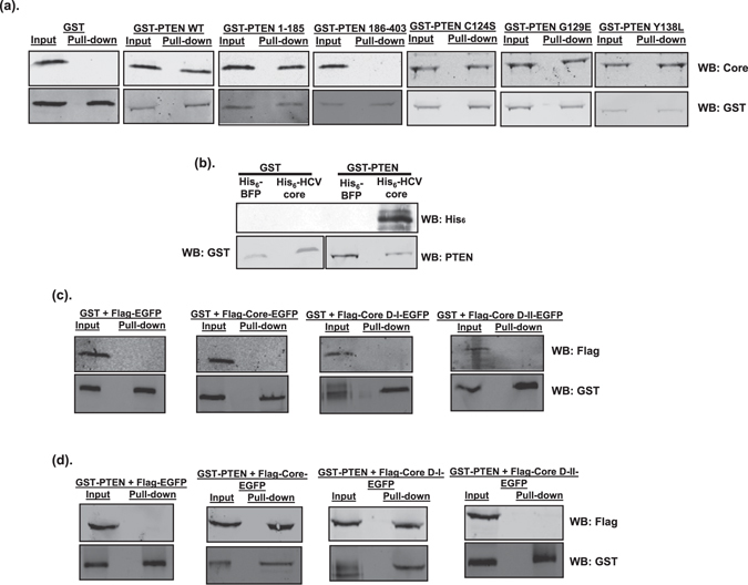
Figure 5.

PTEN interacts with the domain I of HCV core. (a) Huh-7 cells harboring HCV-2a J6/JFH-1(p7-rLuc-2A) replicon were transfected with plasmids expressing GST, GST-PTEN wild-type or GST-PTEN mutants. At 48 hours after transfection, cell lysates were subjected to GST pull-down assay. Input and pull-down products were analyzed by Western blotting (WB) using anti-HCV core and anti-GST antibodies, respectively. (b) Purified His6-BFP (blue fluorescent protein), His6-HCV core, GST and GST-PTEN proteins were subjected to GST pull-down assay. Pull-down products were analyzed by Western blotting (WB) using anti-His6, anti-GST and anti-PTEN antibodies, respectively. Western blot image with a “shorter exposure” can be found in the Supplementary Information. (c and d) Huh-7 cells were co-transfected with plasmids expressing Flag-tagged HCV core, either full-length, domain I (D-I; aa. 1–118), or domain II (D-II, aa. 119–178), together with plasmids expressing GST (c) or GST-PTEN (d). The core proteins were expressed as fusion proteins with EGFP. Flag-tagged EGFP-expressing plasmid was included as negative control. At 48 hours after transfection, cell lysates were subjected to GST pull-down assay. Input and pull-down products were analyzed by Western blotting (WB) using anti-Flag and anti-GST antibodies, respectively.
