
Figure 1.
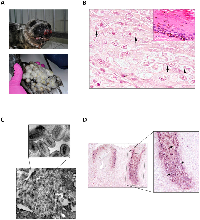
Macroscopic (A), electronmicroscopical (B) and histological analysis (C) of a harbor seal infected with seal parapoxvirus. (A) Ulcerative nodular skin lesion were identified on both front fins and the muzzle of the animal. (B) Histological changes of hair follicle epithelium characterized by ballooning degeneration and cytoplasmic eosinophilic inclusion bodies (arrows), magnification 600x. Normal hair follicle epithelium of an unaffected grey seal is shown in the upper right corner, HE, magnification 600x. (C) Electron microscopy pictures of the cytoplasm of a hair follicle epithelial cell isolated from an infected seal skin lesion. Magnification 37,500x. Mature and immature virus like particles were densely packed. The ovoid to lancealate shape of the core virions are clearly visible. (D) In situ hybridization using a digoxigenin-labeled parapoxvirus-specific DNA probe: parapox virus specific signal is detected in hair follicle epithelial cells of the suprabasal layers (arrow), while no specific signal was detected in the basal layers. Left panel represents an overview with a 100 x magnification, right panel shows a blown up of the highlighted area, magnification: 200x.