
Figure 1.
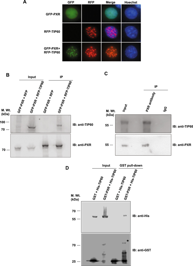
TIP60 modulates intranuclear dynamics of PXR and physically interacts with it. (A) Cos-1 cells were transfected with GFP-PXR and RFP-TIP60 plasmids alone or together and live cell imaging was performed to monitor their subcellular localization. (B) Cos-1 cells were transfected with GFP-PXR and RFP-TIP60 or RFP plasmids. Anti-PXR antibody was used for immunoprecipitation and immunoprecipitated proteins were resolved by SDS-PAGE. Immunoblotting was performed using anti-PXR or anti-TIP60 antibodies as indicated. Input lanes contain 10% of lysate used for immunoprecipitation. (C) To investigate endogenous interaction of TIP60 and PXR, endogenous PXR was immunoprecipitated from HepG2 cells using anti-PXR or control mouse IgG antibodies. Bound proteins were resolved by SDS-PAGE followed by immunoblotting with anti-PXR or anti-TIP60 antibodies. (D) TIP60 and PXR directly interact with each other. His-TIP60 and GST-PXR or GST alone were cotransformed and pull-down of glutathione-agarose-bound proteins was performed. Samples were resolved and immunoblotting was performed using anti-His or anti-GST antibodies. Asterisk indicates full-length band of GST-PXR protein. Full-length immunoblots are presented in Supplementary Figure S6A–C.