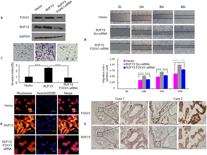
Figure 4.

RUFY3 cooperates with FOXK1 to promote the migration and invasion of CRC cells. (A) Stable transfectants with vector, pooled stable transfectants with RUFY3 transfected with FOXK1 siRNA, and RUFY3 expression were detected by western blot. (B) For the wound healing experiments, cells were analyzed with live-cell microscopy. Original magnification, 10x. ***P < 0.001. (C) Invasive potential of LoVo/Vector, LoVo/RUFY3, and LoVo/RUFY3-FOXK1 siRNA. ***P < 0.001. (D) LoVo cells stained with rhodamine-phallotoxin for 48 h to identify F-actin filaments were visualized under fluorescent microscopy. (E) Representative IHC images are shown for RUFY3 and FOXK1 expression in lymph node metastatic cancer tissues. Scale bars represent 20 μm in D and 100 μm in E. All experiments were repeated three times with similar findings. The full-length blots/gels are presented in Supplementary Figure 8.
