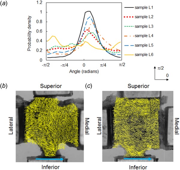
Fig. 4.

Collagen fiber orientation in lumbar FCLs. Fiber orientation distribution (a) and representative fiber alignment maps (for samples L2 (b) and L5 (c)) show that the dominant fiber alignment is along the medial–lateral direction with varied local orientations. The single line arrows indicate the across-joint direction. The orientation distribution functions in (a) were computed relative to the horizontal direction marked by 0.
