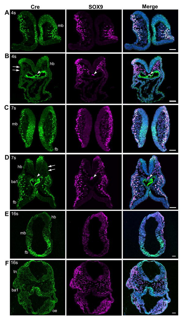
Fig. 10.
Photomicrographs from sections of E8.5 Wnt1-Cre embryos at different somite stages immunostained for the Cre and NC cell marker SOX9. At the 4–7 somite stages (A–D), Cre immunosignals (green) were bright in the midbrain (A, C) and derived NC cells and co-localized with SOX9 (purple). In contrast, in the hindbrain-forebrain levels (B, D) Cre immunosignals were seen in SOX9+ pre-migratory NC cells but were absent in migrating NC cells. In 16-somite embryos (E, F), Cre immunosignals were seen in migrating NC cells in the midbrain and forebrain regions (E). In the trigeminal NC regions (tn), first branchial arch 1 (ba1), and optic eminence region (oe), Cre immunosignals were sparse in migratory NC cells. Scale bars: 50 μm (single-plane laser scanning confocal).

