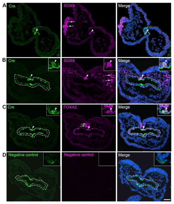
Fig. 3.
Single-plane laser scanning confocal photomicrographs of transverse sections of a 4-somite P0-Cre embryo. Sections were immunostained using an antibody against Cre (green) and were double labeled with SOX9 (purple, A–B) or FOXA2 (purple, C). Arrowheads point to Cre immunoreactive cells co-labeled with SOX9 (A–B) or FOXA2 (C) in the notochord region. Arrows point to single labeled SOX9+ cells in the NC cell region (A–B) and FOXA2+ cells in the floor plate of neural tube (C). White dashed lines outline the foregut diverticulum (B–D). In the negative control slide (D), primary antibodies were omitted. Scale bar: 50 μm for all images.

