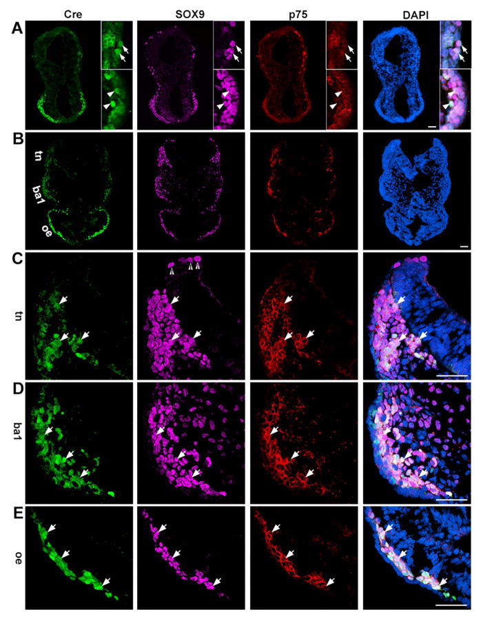
Fig. 5.
Photomicrographs of transverse sections of a P0-Cre embryo at the 14-somite stage. Sections were immunostained for Cre (green), SOX9 (purple), and p75 (red) at the anterior hindbrain-forebrain level (A) and posterior hindbrain-forebrain level (B). C–D are higher magnification images of trigeminal NC (C, tn), branchial arch 1 (D, ba1), and optic eminence (D, oe). Arrows (A) point to Cre+ cells that were co-labeled with SOX9 and p75 immunosignals in the anterior hindbrain regions, and arrowheads (A) point to triple labeled Cre+SOX9+p75+ cells in the forebrain regions. Open arrowheads (C) point to SOX9+ cells in the neural fold region, presumably pre-migratory NC cells. Arrows (C–E) point to Cre+ cells co-labeled with SOX9+ and p75+, presumably migratory NC, in trigeminal NC regions (tn, C), first branchial arch 1 (ba1, D) and optic eminence (oe, E). Scale bars: 50 μm for all images (single-plane laser scanning confocal).

