Fig. 8.
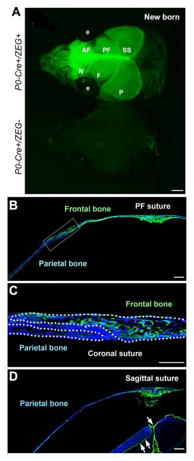
P0-Cre marks cranial NC and derived cells in the skull. A: GFP signals in the newborn P0-Cre+/ZEG+ (upper) and control littermate (bottom) mice. B: GFP signals on the coronal tissue sections illustrate that the frontal bones (B–C), but not the parietal bone (C–D), were labeled. C is the high magnification image of the squared tissue region in B. Abbreviations: AF, anterior frontal suture; e, eye; F, frontal bone; N, nasal bone; P, parietal bone; PF, posterior frontal suture; SS, sagittal suture. Scale bars: 500 μm in A, 200 μm in B and D; 100 μm in C.

