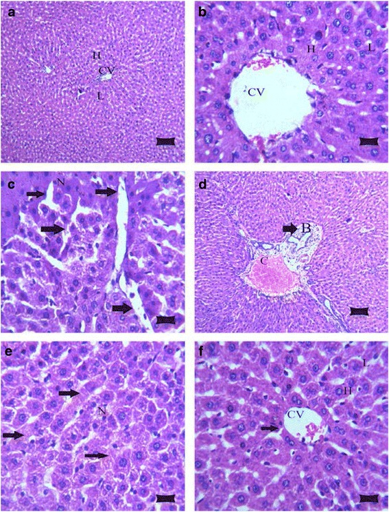
Fig. 1.

Photomicrograph of rat livers in the various groups: a Control group, showing the normal histological structure of hepatic lobule (L) with centrally located euchromatic nucleus of hepatocyte (H) surround central vein (CV) (H&E; bar, 89 μm); b Karela administered group, shows normal histological structure of hepatic lobule (L), hepatocyte (H) and central vein (CV) (H&E; bar, 14 μm); c Cholesterol group, showing deposition of cholesterol crystals in many radicular cysts forming cholesterol clefts (arrows) and necrosis (N) of hepatocytes (H&E; bar, 14 μm); d Cholesterol administered group, showing sever congestion of portal blood vessel (C) with extensive portal fibrosis (arrow) and hyperplasia of epithelial lining of the bile duct (B) (H&E; bar, 89 μm); e Cholesterol group, showing necrosis in hepatocytes (N) with karyolysis of hepatocytes nuclei (arrows) (H&E; bar, 14 μm); and f Cholesterol and Karela group, show apparent normal hepatic parenchyma with few cholesterol clefts (arrow), hepatic lobule (L), hepatocyte (H), central vein (CV) (H&E; bar, 14 μm)
