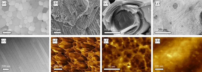
Figure 2.
Diverse microstructures and universal nanostructures in biominerals. (a) Optical micrographs of the prismatic layer of P. nobilis. (b) Electron microscopical image of crossed-lamellae in G. glycymeris. (c) STEM images of a nacreous layer in P. nobilis. (d) Atomic force microscopy reveals, here in the phase image, the nanogranules of which the prisms of P. nobilis consist. (e) The glass sponge Euplectella sp. also consists of similar granules; the phase image in (f) reveals that the silica grains are of smaller diameter as in its calcareous counterparts. (g) Bone also consists of nanoscale particles; here, the bone apatite nanoparticles are also distinctly smaller, as revealed by the height image (h).

