Fig. 6.
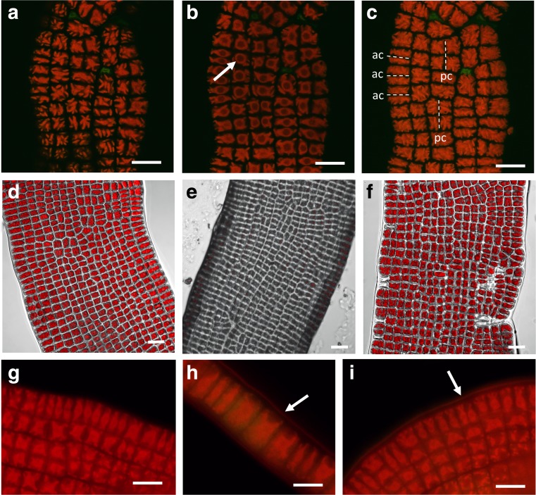
Confocal laser scanning microscopy (a–f) and epifluorescence microscopy (g–i) of Prasiola calophylla. a Cortical optical section of chloroplast lobes. b Median optical section showing the round pyrenoids free of chlorophyll autofluorescence. c z-stack projection allowing to depict the division planes of the cells. ac anticlinal cell division, pc periclinal cell division. d Freshly harvested thallus segment illustrating the cell walls and the chloroplast autofluorescence. e Segment of the same thallus desiccated for 2.5 h at 20% RH; chlorophyll autofluorescence (false color red) declined strongly. f Segment of the thallus rehydrated for 1.5 h. g Control thallus segment where nile red staining was omitted. h Uniserate filament stained with 0.1% nile red. i Thallus segment stained with 0.1% nile red, showing reddish fluorescence labelling on the surface of the thallus (arrows). Bars 10 μm

