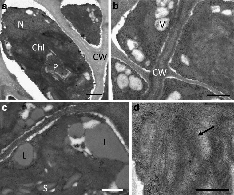
Fig. 8.
Transmission electron micrographs of filed-collected Prasiola calophylla samples desiccated at ∼20% RH for 2.5 h. a Dense cytoplasm slightly detached from the cell wall, pyrenoid clearly visible. b Cells with homogenous cell content, small vacuoles in the cell cortex. c Detail of a cell containing large lipid bodies and starch grains. d Detail of the chloroplast with still intact thylakoid membranes (arrow). Chl chloroplast, CW cell wall, L lipid body, N nucleus, P pyrenoid, S starch grain. Bars a–c 1 μm, d 0.5 μm

