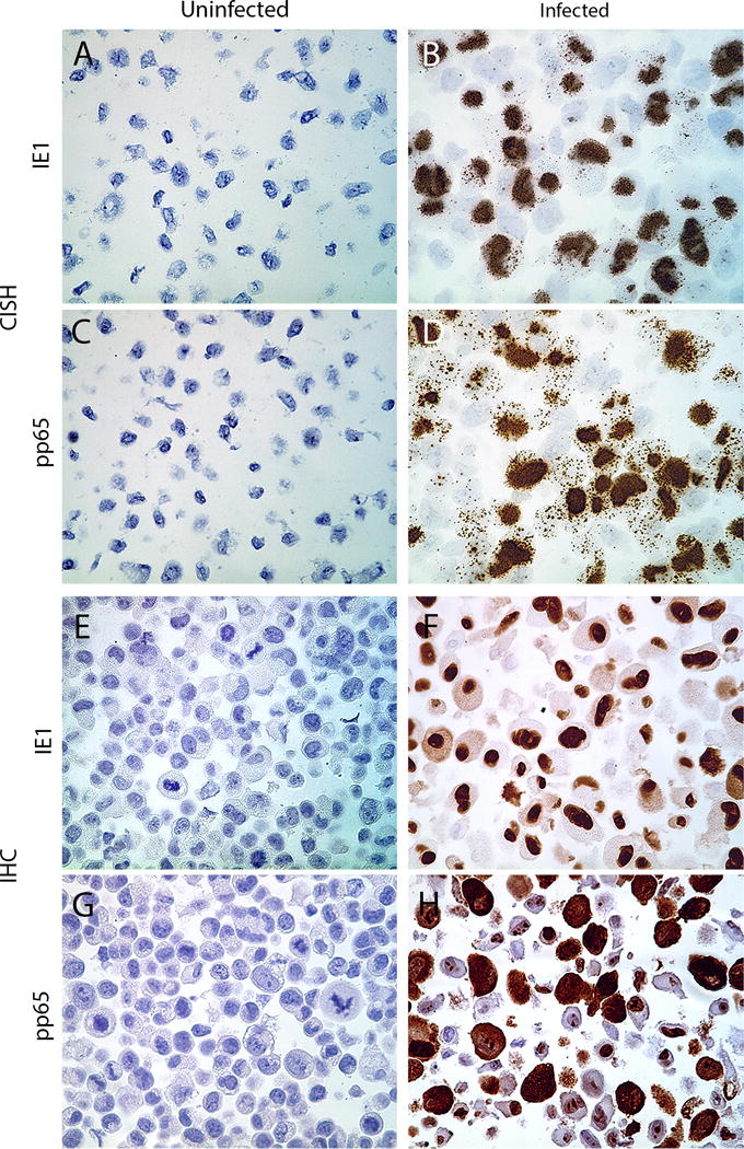
Figure 1. Analytical validation of CISH and IHC in human fibroblast cell lines.

Human foreskin fibroblasts were uninfected or infected with a human CMV Towne for 72 h. Cells were then fixed in neutral buffered formalin overnight and processed into paraffin blocks. CISH assay depicts targeting IE1 DNA in uninfected (A) and CMV-infected (B), and pp65 DNA in uninfected (C) and CMV-infected cells (D). Representative figures for IHC staining of IE1- and pp65 proteins in uninfected (E,G) and CMV-infected (F,H) cells are shown (all images are original magnification of ×400).
