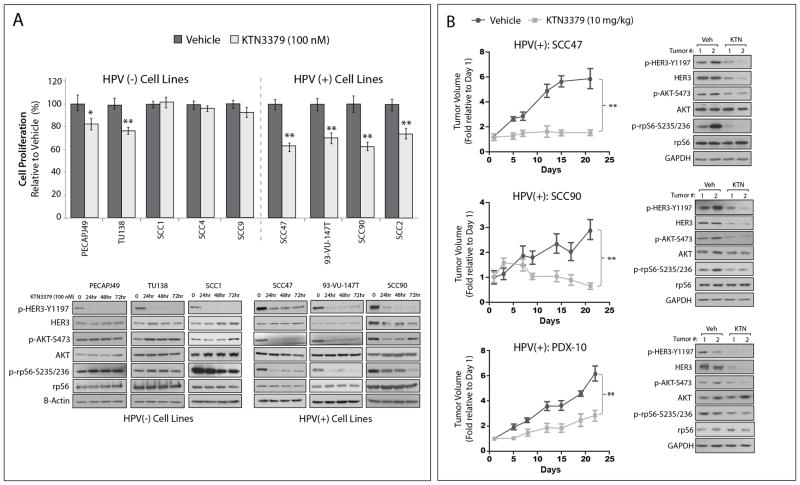
Figure 5. HPV(+) HNSCC models are sensitive to therapeutic blockade of HER3 with the anti-HER3 monoclonal antibody, KTN3379.
(A) HPV(−) and HPV(+) HNSCC cell lines were treated with 100 nM KTN3379 for 72 hours before performing proliferation assays. Proliferation is plotted as a percentage of growth relative to vehicle treated cells (n=6 in three-five independent experiments). Data points are represented as mean ± s.e.m. *, P <0.05, **, P <0.01. Whole cell lysates were harvested from three HPV(−) cell lines (PECAPJ49, TU138, and SCC1), and three HPV(+) cell lines (SCC47, 93-VU-147T, and SCC90) over a time course of 72 hours of KTN3379 treatment (100 nM) followed by immunoblotting for the indicated proteins. β-actin was used as a loading control. (B) HPV(+) cell lines (SCC47 and SCC90) were grown as xenografts in athymic nude mice (n=6 tumors per treatment group for SCC47, and n=4 tumors per treatment group for SCC90). The HPV(+) PDX-10 was implanted into the dorsal flanks of NOD/SCIDγ mice (n=6 tumors per treatment group). All xenografts were treated with KTN3379 (10 mg/kg) or vehicle twice weekly for the indicated time period by IP injection. Tumor volumes for all xenograft experiments were calculated as fractions of the average starting volume for each group, and the mean tumor volume ± s.e.m is shown. **, P<0.01.

