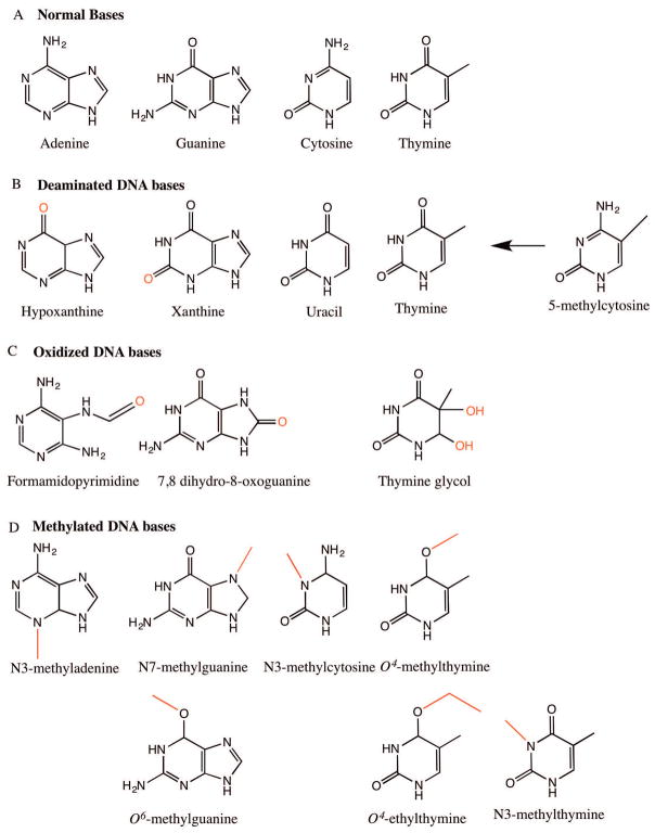
Figure 1.
Common DNA base lesions. A) Normal structures of DNA bases: adenine (A), guanine (G), cytosine (C) and thymine (T). B) Deaminated bases: hypoxanthine, xanthine, uracil and thymine arising from deamination of exocyclic bases of adenine, guanine, cytosine and 5-methylcytosine (5-mC) respectively. C) Oxidized DNA bases: formamidopyrimidine derivative of adenine (Fapy-A), 7,8 dihydro-8-oxoguanine (8-oxo-G) and thymine glycol. D) Methylated DNA bases: N3-methyladenine, N7-methylguanine, O6-methylguanine, N3-methylcytosine, O4-methylthymine, O4-ethylthymine and N3-methylthymine.

