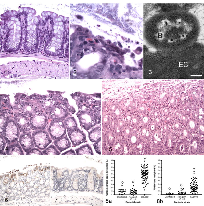
Figure 1.

Uninfected germ-free control mouse, normal colon. Hematoxylin and eosin (HE). Figures 2–6. Enterohemorrhagic Escherichia coli (EHEC)-monocolonized mice, colon. Figure 2. A focus of colon epithelial necrosis associated with large numbers of adherent bacteria. Short bacilli adhere to both superficial and sloughed epithelial cells. HE. Figure 3. There is an adhesion-pedestal-like structure and close association between a degenerate epithelial cell (EC) and a bacterium (B). Bar = 250 nM. Transmission electron microscopy. Figure 4. Severe colonic epithelial necrosis and apoptosis compared to normal colon from a germ-free mouse (see Fig. 1). HE. Figure 5. Necrosis and sloughing of necrotic cells into the lumen, and neutrophilic infiltration in the lamina propria. HE. Figure 6. Activated caspase-3 immunoreactivity in the colonic epithelium is characterized by brown granular staining in surface cells, extending into the glandular epithelial cells, and in sloughed cells within the lumen. Immunoperoxidase-DAB. Figure 7. Uninfected germ-free mouse, colon. Caspase-3 immunoreactivity is not detectable in epithelial cells. Immunoperoxidase-DAB. Figure 8. Colonic epithelial necrosis (a) and neutrophil infiltration scores (PMN score) (b) in uninfected germ-free mice and in gnotobiotic mice monocolonized with nonpathogenic E. coli or with EHEC strain EDL933 and euthanized 1 week after bacterial colonization. *Significantly different from uninfected. ◊Significantly different from EDL933.
