
Figures 9–11.
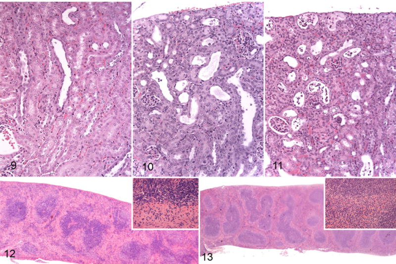
Enterohemorrhagic Escherichia coli (EHEC)-monocolonized mice, tubular necrosis, kidney. Hematoxylin and eosin (HE). Figure 9. Tubular necrosis score 1. Few dilated tubules with epithelial atrophy and rare sloughed cells. Figure 10. Score 2. Several dilated tubules with epithelial atrophy and regeneration, luminal protein, and sloughed epithelial cells. Figure 11. Score 3. Most tubules are dilated with flattened epithelium and contain sloughed epithelial cells. Figure 12. EHEC-monocolonized mouse, spleen. There is marked loss of cellularity in the red pulp. Lymphoid tissue is mildly depleted. Inset: junction between red pulp (bottom) and white pulp (top) with marked loss of hematopoietic cells and scattered karyorrhexis in the red pulp and scattered apoptosis in the white pulp. HE. Figure 13. Uninfected germ-free mouse, normal spleen. Inset: junction of red pulp (bottom) and white pulp (top). HE.