
Figure 7. Human Melanoma Granzyme B Analysis and Peptide Binding.
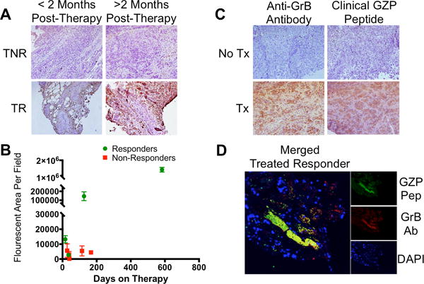
A) Human melanoma samples from patients treated with an anti-PD-1 checkpoint inhibitor were grouped as treated responders (TR) or treated non-responders (TNR) based on modified RECIST criteria. B) Immunofluorescent microscopy quantification of granzyme B in 9 patients at incremental time intervals reveals significant differences in granzyme B expression as early as 16 days post-therapy between responding and non-responding patients. C) Comparison of matched samples using either an anti-Granzyme B antibody (Anti-GrB) or humanized GZP peptide (clinical GZP) reveals similar patterns of staining between the antibody and peptide that are much stronger in treated patients than untreated patients. D) Co-localization (yellow) of human GZP (green) and anti-GrB (red) are demonstrated in a treated responder melanoma patient, with nuclear staining (DAPI) in blue.