
Figure 6. STAT1 Is a Key Factor in the Signaling Pathway that Connects CD95 to Cancer Stemness.
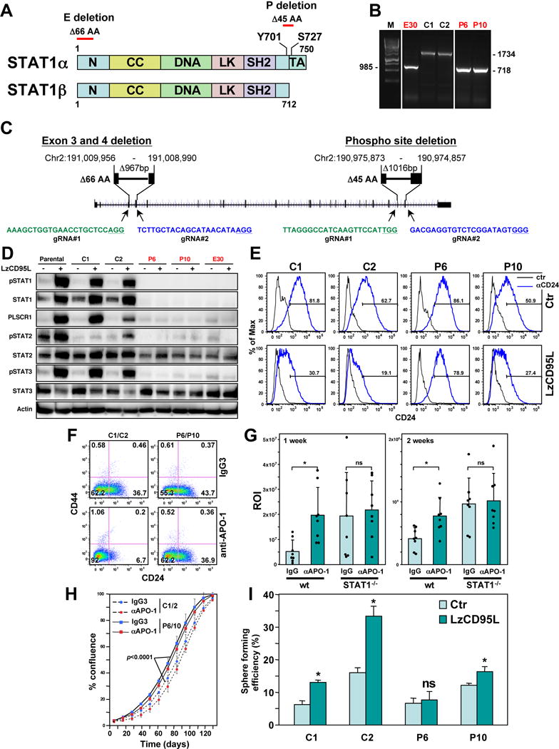
(A) Schematic of the STAT1 protein and the two sites deleted by CRISPR/Cas9 gene editing.
(B) PCR confirmation of homozygous deletions at the two sites in STAT1 in three isolated clones (labeled in red).
(C) Genomic localization of the two CRIPSR/Cas9 deletions in the human STAT1 gene (Human Dec. 2013 (GRCh38/hg38) assembly). The PAM sites in the guide (g)RNAs are underlined. gRNAs in green target the sense and in blue the antisense strand.
(D) Western blot analysis of MCF-7 cells, Cas9 transfected wt clones and STAT1 k.o. clones either control treated or treated with LzCD95L for 4 days.
(E) CD24 surface staining of the clones treated as in D.
(F) CD44/CD24 surface staining of cell pools of clones C1 and C2 and P6 and P10 (now expressing a luciferase plasmid for the in vivo experiment in H) after 7 days of treatment with either IgG3 or anti-APO-1.
(G) Bioluminescence of tumors in NSG mice one week and two weeks after injection of the cells analyzed in F into the fat pad.
(H) Growth of the pooled cells after 9 days of treatment with either IgG3 or anti-APO-1. p-value was calculated using ANOVA.
(I) Percent of wells with spheres after plating MCF-7 cells (treated with LzCD95L for 6 days) at one cell per well in 96-well plates. p-value *<0.05; ns, not significant.
See also Figures S7–S9.