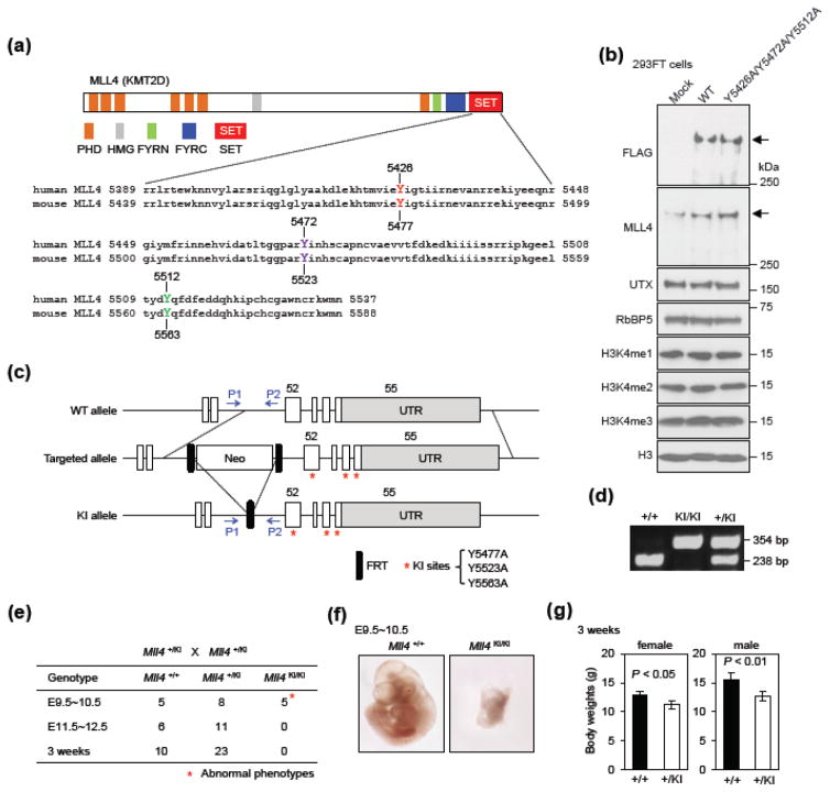
Fig. 2. MLL4 enzymatic activity is essential for early embryonic development.
(a) Upper panel shows the domains in human and mouse MLL4 proteins. Lower panel shows the alignment of amino acids in the C-terminal SET domain of human and mouse MLL4. Tyrosine (Y) residues that are potentially critical for enzymatic activity are highlighted in color. (b) 293FT cells were transfected with pCMV6-MLL4 plasmid expressing FLAG-tagged WT human MLL4 or the Y5426A/Y5472A/Y5512A mutant. Nuclear extracts and histone extracts were analyzed by Western blot using antibodies indicated on the left. The arrows indicate the expected position of MLL4 protein. (c) Schematic representation of mouse Mll4 WT, targeted and enzyme-dead knock-in (KI) alleles. Deletion of neo selection cassette from the targeted allele by FLP recombinase generates the KI allele. The KI allele carries Y5477A/Y5523A/Y5563A, represented by the red asterisks. The locations of PCR genotyping primers P1 and P2 are indicated by arrows. (d) PCR genotyping of ES cell lines using P1 and P2 primers. (e) Genotyping results from crossing Mll4+/KI with Mll4+/KI mice. The expected ratio of the three genotypes is 1:2:1. (f) Representative images of E9.5~10.5 embryos. (g) Body weights of 3-week-old Mll4+/KI and Mll4+/+ mice (n = 3 for female and n = 6 for male). Data are presented as mean ± SD and P value by Student’s t-test.

