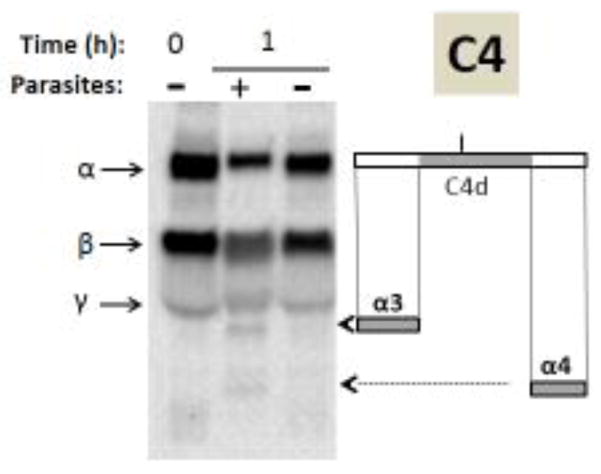
Figure 5.
Analysis of complement protein C4 by western blotting. Samples obtained following human serum incubation for 1 hour at 37°C either in the presence of schistosome parasites (+) or without parasites (−) taken at 0 or 1 hour (as indicated) were resolved by SDS-PAGE, blotted to PVDF and probed with anti-human C4 antiserum. The arrows indicate the α, β and γ chains of C4 and the arrowheads indicate the positions of migration of the C4α breakdown products α3 and α4. At right is a depiction of the C4α chain (large rectangle) and beneath this the α3 and α4 fragments are indicated (small grey rectangles). The central region of C4α represents the C4d fragment (colored grey in upper rectangle) and the short vertical bar above C4d indicates the position of molecular attachment of C4 to pathogens once complement is engaged.

