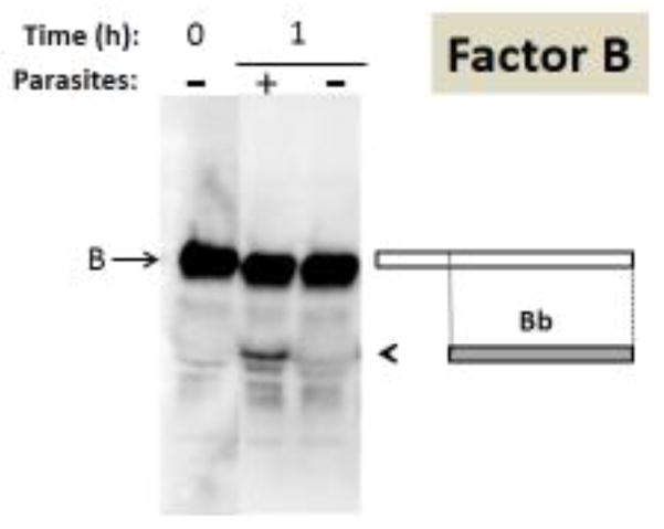
Figure 6.
Analysis of complement protein factor B by western blotting. Samples obtained following human serum incubation for 1 hour at 37°C either in the presence of schistosome parasites (+) or without parasites (−) taken at 0 or 1 hour (as indicated) were resolved by SDS-PAGE, blotted to PVDF and probed with anti-human factor B antiserum. The arrow indicates factor B and the arrowhead indicates the position of migration of the large fragment of factor B designated Bb. At right is a depiction of factor B (large rectangle) and beneath this the Bb fragment is indicated (smaller grey rectangle).

