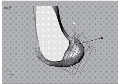
Figure 4. Lateral view of the medial femoral condyle. Red dot indicates mean center of ACL tunnel position using Rhinoceros. Blue dot represents the same position using OsiriX. Note the difference in height (H) between radiological methods. Distance between tunnels (d) = 2.02 mm.

