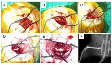
Figure 1. (A) An image of the femur after retraction of the soft tissues; (B) After the transverse fracture was obtained; (C) After placing an anterograde intramedullary K-wire from the fracture site to the knee; (D) Retrograde movement of the K-wire after reducing the fracture; (E) The last image of the fracture after pushing the K-wire inside the bone; (F) The postoperative roentgenogram of the fracture.

