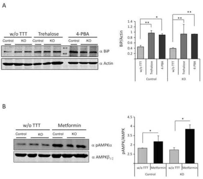
Figure 1.

Drugs used in this study affect their corresponding targets in mice brain. Western blot analyses of whole brain extracts using the indicated antibodies and densitometric quantification of the corresponding blots were carried out as described in Material and Methods. A representative blot is presented. (A) anti-BiP/GRP78 (BiP) and anti-actin (loading control) from Epm2b+/− (control) and Epm2b−/− (KO) mice untreated (w/o TTT) or treated with trehalose (2%) or 4-PBA (20 mM) for two months. *p<0.05, **p<0.01 (n: 3 [control] or n:4 [KO]) comparing the indicated groups with the basal condition according to Kruskal-Wallis non-parametric test followed by Conover-Inman post-hoc test. (B) anti-pThr172-AMPKalpha (pAMPKα) and anti-AMPKβ1/2 (loading control) from Epm2b+/− control and Epm2b−/− (KO) mice untreated (w/o TTT) or treated with 12 mM metformin for two months. Bars indicate mean values of the relative intensity of each protein of at least three independent samples ± SEM. Asterisks denote significant differences *p<0.05, (n: 3 [control] or n:4 [KO]) comparing the indicated groups with the basal condition according to the Wilcoxon-Mann-Whitney test.
