
FIGURE 2.
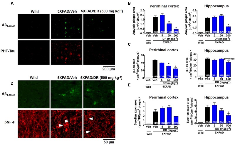
DR extract ameliorates AD-like pathology in 5XFAD mice. DR extract (5, 50, 500 mg kg-1, p.o.) or vehicle solution (saline) was administered to wild-type or 5XFAD mice (males and females, 6–8 months old) for 31 days. The day after the behavioral test, mice were sacrificed and subjected to brain analysis. (A) Representative images of Aβ1-40/42-positive plaques and PHF-tau in the perirhinal cortex. (B) The total area of amyloid plaques per 100 μm2 was quantified in the cerebral cortex and hippocampus. (C) The total area of PHF-tau associated with Aβ plaques per 100 μm2 was quantified in the cerebral cortex and hippocampus. (D) Aβ plaques and axons were double immunostained with Aβ1-40/42 and pNF-H antibodies. pNF-H-positive bulb- or ring-like axonal structures were localized with Aβ plaques. Representative images from the perirhinal cortex are shown. (E) The total area of abnormal axons per 100 μm2 of amyloid plaque in the cerebral cortex and hippocampus are shown. One-way ANOVA and Dunnett’s post hoc test were used for all statistical analyses (∗p < 0.05 vs. veh/5XFAD; the number of measured areas is shown in each column).