Figure 1.
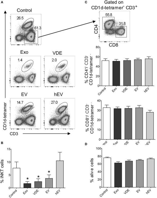
Invariant natural killer T (iNKT) cell expansion was impaired in the presence of Leishmania infantum Exo, extracellular vesicle (EV), and vesicle-depleted-exoproducts (VDEs). (A) Representative FACS profile showing the gating strategy used to identify iNKT cells following 8–12 days of culture with α-GalCer alone (control) or together with L. infantum Exo, EV, VDE, or hEV. iNKT cells were identified as CD3+ CD1d-tetramer+ cells. (B) Histograms represent the percentage of iNKT cells, as described in (A). (C) Representative FACS profile showing the gating strategy used to identify CD4+ and CD8+ conventional T cells among CD3+CD1d tetramer− peripheral blood mononuclear cell (PBMC) after 8–12 days of culture in the presence of α-GalCer. Histograms represent the percentage of CD4+ and CD8+ T cells among CD3+CD1d tetramer− cells. (D). Histograms represent the percentage of living cells, detected by 7AAD staining, among whole PBMC cultured in the presence of α-GalCer. Data are shown as means ± SEM (n = 14–24 individual donors tested). All groups were tested versus control group. *p < 0.05.

